How extreme dieting can affect bone health

In a recent Instagram post, the actor Jameela Jamil revealed she has poor bone density, despite only being in her 30s. Jamil blamed this finding on 20 years of dieting – urging her followers to be aware of the harms diet culture can do to your health.

Bone density is important for many reasons, primarily because it acts as a reservoir for many of the important minerals our bones need to function well. Many factors can affect your bone density – and as Jamil has pointed out, diet is one component that has a significant effect on bone health.

Bone is a living tissue. This means our skeleton grows and remodels itself according to the stresses and strains it’s put under. Everything from fractures to exercise require our bones to change their shape or density. This is why a weightlifter’s skeleton is much denser than a marathon runner’s.

The biggest skeletal changes we experience happen in our younger years. But bones keep changing throughout our lives depending on how active we are, what our diet consists of, and if we’ve suffered an injury or disease.

Bones are made of proteins, such as collagen, as well as minerals – largely calcium. This is a key mineral for us, as it keeps our bones and teeth strong and helps repair and rebuild any injured bones.

But other minerals and vitamins are also important. For example, vitamin D supports calcium, playing a key role in bone mineralisation. This is where calcium combines with phosphate in our bones to create the mineral crystal hydroxyapatite. This crystal is crucial to our bone mineral density (also known as “bone mass”), as it helps bones remodel and maintain their structural strength.

Dexa scans – the type of scan Jamil referred to in her post – can measure the density of these crystals in bones. The more hydroxyapatite crystals detected, the healthier the bones are.

The more crystals detected, the better your bone density.
Crevis/ Shutterstock

We hit peak bone mineral density in our late teens and early 20s, when our body has grown to full size and our metabolism is working its best. From here, it’s possible to maintain stable bone mass into your late 30s for women and early 40s for men, with the right diet and activity. But after this point, it begins to decline.

Bone density

We accrue calcium over many years. It initially comes from our mother, then later from our diet. Our body accrues calcium so it can adapt to times when calcium demand is greater than what we can get from our diet – such as during pregnancy, when the foetus needs calcium to build its own bones.

However, relying solely on this skeletal calcium reserve can’t be sustained for lengthy or repeated periods, because of how long it takes to be replenished. This is why diet is so important for bone density – and why a poor diet can cause extreme damage, especially when certain food groups or minerals are consistently left out.

For instance, studies have shown consuming soft drinks, (particularly cola), more than four times a week is linked with lower bone density and increased fracture risk. This is true even after adjusting for many other variables that affect bone density.

These carbonated and energy drinks contain varying levels of vitamins – often with none of the minerals, including calcium, that the body needs to function optimally. This causes the body to draw on its reserves if calcium isn’t being delivered elsewhere in the diet.

Diets high in added sugar can also have a detrimental affect on the skeleton. Excess sugar causes inflammation and other physiological changes, such as obesity. Consuming high amounts of sugar is linked with reduced calcium intake, especially in children who substitute milk for sugary drinks. Excess sugar consumption also causes the body to excrete excess calcium, instead of reabsorbing it in the kidney as the body normally would.

Low- and high-fat diets have also been associated with increased risk of osteoporosis (a condition that weakens bones) in women – though larger studies are needed to better understand the effects of removing whole food groups on bone health.

Anorexia nervosa also has a significant affect on bone density – affecting a majority of people with the condition.

Low bone mineral density – especially in the spine – puts people with anorexia at increased risk of fractures because their bone thickness is reduced, increasing the likelihood of developing osteoporosis, which is associated with increased fractures.

Anorexia in young adulthood is particularly challenging. This is the stage where the skeleton is building itself to reach peak bone mass, so it’s depositing calcium at a record pace. When diet is insufficient and the body already starts drawing on its mineral reserves, there’s a potential that the bone density or calcium reserves in the body will never be optimal – increasing fracture risk for the rest of that person’s life.

Can bone health be fixed?

Optimal bone health starts in utero, but our prepubescent years are key to setting our skeleton up for later life. People who are behind the curve in early life may have difficulty achieving their peak, as poor bone mineral density can affect everything from our appetite to how efficient our gastrointestinal tract is at absorbing important nutrients (including calcium). Supplements have a limited effect because our body can only absorb a set amount of any vitamin or mineral at a time.

While it’s possible to limit some of the decline in bone density that naturally happens as we age, some of the choices we make – such as not consuming enough calcium – can accelerate the decline. Biological sex also has a significant impact on our bone health in old age – with post-menopausal women at greater risk of osteoporosis because they produce less oestrogen, which helps keep the cells that degrade bone in check. Läs mer…
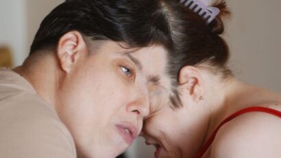
The world’s oldest conjoined twins have died – what we know about this rare condition

The world’s oldest conjoined twins, Lori and George Schappell, recently died, aged 62.

Doctors predicted that the twins (who were joined at the skull and shared 30% of their brain) would not live past the age of 30. But the twins defied expectations and managed to live long and successful lives.

The twins made headlines in 2007 when George came out as transgender.

Conjoined twins are incredibly rare, accounting for about one or two in every 100,000 births. Sadly, about 60% of conjoined twin births are stillborn or die shortly after birth. Conjoined female twins are three times more likely to survive than males.

Although all conjoined twins are identical (monozygotic), recent genetic analyses have revealed that there are differences in genes, including those linked to growth and development.

Because conjoined twins are so rare, it is difficult to know exactly how they come about. However, there are two possible causes, both of which have some evidence to support them, but neither can completely account for the full range of presentations that are seen.

The first process is “fission”, where a single fertilised egg does not fully split during the process of forming identical twins. The second process is “fusion”, where two fertilised eggs fuse soon after fertilisation occurs.

Types of conjoined twins

There are 15 recognised types of conjoined twins, based on the various places their bodies fuse.

The most common fusion site is the chest and abdomen (thoraco-omphalopagus). Other common fusion sites are the chest (thoracopagus), the abdomen (omphalopagus) and the skull (craniopagus).

The first part of the word is the body part that is fused, and “pagus” is Greek for “fixed”. They can also be described by number (bi, tri or tetra for two, three or four), so a “dicephalic parapagus” conjoined twin would have one trunk and two heads.

Conjoined twins are usually picked up on routine ultrasound scans performed during pregnancy. This would then be followed up by MRI scans to get a clearer picture of the type of fusion as well as information on which organs are shared. This helps healthcare professionals to manage any complications that occur during delivery and provides useful information on potential surgical separation further down the line. Sadly, it also provides information on whether a termination is necessary.

Conjoined twins who are diagnosed by imaging are typically delivered by caesarean section.

George enjoyed a successful career as a country singer while Lori was a trophy-winning ten-pin bowler.
Associated Press/Alamy Stock Photo

Separating twins

Surgery to separate conjoined twins is highly complex and only a few hospitals and surgical teams have performed them; they include Great Ormond Street Hospital in London, Children’s Hospital of Philadelphia and Red Cross Children’s Hospital in Cape Town. Surgeons in Saudi Arabia are also renowned for performing these surgeries, with the surgeon to the Royal Court performing at least 60.

As medical advances continue, more hospitals are beginning to undertake these operations, which can last more than 24 hours. One of the longest operations to separate conjoined twins, who were fused at the cranium, took more than 100 hours.

The earliest recorded attempt at surgical separation of conjoined twins dates back to AD945 in Armenia where conjoined brothers lived until middle age before one of them died. Surgeons tried to separate the living brother from his deceased sibling, but he died a couple of days later.

The first successful surgical separation of conjoined twins was undertaken in 1689. These were xiphophagus twins, meaning they were fused by the bottom of their sternums. In slightly more recent history (1860), a surgeon separated his own omphalopagus daughters. Unfortunately, only one survived.

Those fused at the abdomen (omphalopagus) have the best chance of surviving separation surgery. There are more than 100 documented cases of both separated twins surviving.

Separating conjoined twins requires a significant amount of planning before surgery can begin, including, of course, lots of imaging such as ultrasound, CT and MRI.

Although this imaging will tell doctors whether each twin has their own set of organs – and which, if any, are connected – it doesn’t tell them whether each can function fully to support the respective twin in an independent life.

This is where further imaging assessments are needed using technology such as fMRI, contrast-enhanced CT, diffusion tensor imaging and vascular imaging to determine if systems such as the liver, gut and urinary systems of each twin have their own blood supply to ensure they can function after they are separated.

While surgery commences as a single operation, as it progresses it becomes two separate surgeries once the twins are separated, which requires two teams of surgeons, anaesthetists and theatre staff to deal with each child.

Twins who share vital organs – or whose organs are fused – have a lower chance of a successful separation.

Krista and Tatiana Hogan, Canadian conjoined twins share a thalamus (an egg-shaped structure in the middle of the brain), which raises questions about whether their experiences can be felt by the other, even though they are separate people.

The process of separation of conjoined twins raises ethical questions about potential harm or even death of one twin to benefit the other, and this is often a significant issue in any potential surgical intervention. Läs mer…

Robert F. Kennedy Jr. reveals why he has a raspy voice – spasmodic dysphonia explained

Anyone following the US presidential race may have noticed independent candidate Robert F. Kennedy Jr. has a particularly harsh-sounding voice. The cause is a neurological condition called spasmodic dysphonia.

Spasmodic dysphonia, also known as laryngeal dysphonia, often results in a shaky, tight or strained-sounding voice. It does not affect other functions of the vocal cords, such as laughing, crying or shouting.

It affects about one in every 50,000 people – with women being more prone to it than men. It usually develops between the ages of 30 and 50.

The causes aren’t clear, but one study found that 65% of people with the condition previously had measles or mumps – compared with the US national average of 15%.

A separate study found the measles and mumps vaccines helped protect against developing the condition. An interesting finding, if confirmed, given that RFK Jr. is a noted vaccine sceptic.

Other studies have suggested that a history of throat and sinus illnesses, mumps and rubella, intense occupational voice use, tremor, tics and compulsive behaviour are all associated with spasmodic dysphonia.

There are three types of spasmodic dysphonia, the most common being “adductor spasmodic dysphonia”, accounting for 85-95% of cases. In this version of the condition, the muscles that bring the vocal cords together are affected, making the cords stiffen or slam shut as a result of the spasms. This causes a strained or strangulated sound during speech.

The less common type is “abductor spasmodic dysphonia”, which causes spasms that trigger the cords to open. The reason this is rarer is there is only one muscle (posterior cricoarytenoid) on each side that opens the cords. This results in speech being quieter or weak.

Finally, incredibly rare is a spasmodic dysphonia that affects both the adductors and abductors.

There is no cure for spasmodic dysphonia, but there are treatments that can help alleviate the symptoms.

Botox injections have become the gold standard therapy. However, this is a short-term solution and requires an injection every three to six months. Speech therapy can also help by strengthening the defective muscles or enabling other muscles to compensate. Sometimes Botox and speech therapy are combined.

A more invasive treatment involves severing some of the adductor muscles (for the more common form of the condition). This mimics a permanent Botox injection.

A recent small study also showed that deep-brain stimulation (a type of pacemaker for the brain) can improve the voice quality of people with adductor spasmodic dysphonia.

RFJ Jr. talks about his raspy voice.

Voice changes can signal other health problems

Ageing causes our voices to change. Over time, our vocal cords become stiffer and less flexible, which alters the sound of our voice. People who use their vocal cords regularly – such as singers – show significantly less change in sound than those who don’t sing.

But voice changes over a shorter period can suggest an illness.

Respiratory illnesses

Our respiratory system is lined by a special layer of cells called “pseudostratified columnar ciliated epithelium”. Among the columnar cells are specialised cells called goblet cells that produce mucus. The role of mucus is to prevent microbes (common cold, COVID and many other things) from getting deep into the respiratory system, by causing microbes to stick.

The cilia then beat the mucus upwards, about ten to 14 times a second, towards the larynx where the microbe-containing-mucus is swallowed and neutralised by stomach acid.

This accumulation of mucus is increased in people with vocal disorders, because the cords move less and so they are less likely to be able to clear the mucus.

Studies have shown that the most efficient way to remove excess mucus is by hard throat clearing.

Acid reflux

Acid reflux can make the voice hoarse. The rising stomach acid can cause swelling and scarring in the larynx. This changes the shape and structure of the cords, affecting their function and the sound they make.

Allergies

Allergies are a significant contributor to vocal changes. The inflammatory reaction caused by exposure and activation by allergens, such as pollen, cause swelling in the vocal cords and increased mucus.

Antihistamines are often used to treat allergies. But, because they reduce the amount of mucus produced, they tend to dry out the larynx, making the voice very raspy.

Cancer

Changes in vocal sounds without other symptoms or identifiable causes may be caused by cancer tumour growth in the chest. The nerve that controls a muscle that abducts (opens) the vocal cords can become compressed by tumours in the chest. This is because it travels down from the skull, into the chest, wrapping under the aorta before returning to supply the larynx.

Studies have shown that between 18% and 24% of people with tumours that are in the chest – but not their larynx – have vocal cord paralysis that causes their voice to change.

Vocal changes happen subtly with ageing, but there are many other causes. Any changes that persist for weeks should be checked by a GP, particularly if there are other symptoms accompanying it. Läs mer…

Eye infections might seem like a minor complaint – but in some cases they can cause blindness and even death

When you think of eye infections, what comes to mind? Puffy, swollen bruised feeling eyelids that get glued together with gunk overnight? That feeling of having grit in your eye that can’t be cleaned away? Eye infections may seem like a relatively minor – if unsightly and inconvenient – complaint, but they can also be far more serious.

Take the deadly outbreak of antibiotic resistant bacteria Burkholderia cepacia in 2023-24, for example.

Between January 2023 and February 2024, contaminated brands of lubricating eye gel were linked to the infection of at least 52 patients. One person died and at least 25 others suffered serious infections.

The outbreak has now subsided and products are back on the shelves but it isn’t the first time that medicinal products have led to outbreaks of B cepacia.

The bacteria is an opportunistic pathogen known to pose a significant risk to people with cystic fibrosis, chronic lung conditions and weakened immune systems. The infection likely progresses from the mucous membranes of the eyelids to the lungs where it leads to pneumonia and septicaemia causing death in days.

But it’s not just B cepacia that can threaten our health. Something as simple as rubbing our eyes can introduce pathogens leading to infection, blindness and, in the worst case, death.

Bacteria account for up to 70% of eye infections and globally over 6 million people have blindness or moderate visual impairment from ocular infection. Contact lens wearers are at increased risk.

The eye is a unique structure. It converts light energy to chemical and then electrical energy, which is transmitted to the brain and converted to a picture. The eye uses about 6 million cones and 120 million rods which detect colour and light.

Eye cells have no ability to regenerate so, once damaged or injured, cannot be repaired or replaced. The body tries its best to preserve the eyes by encasing them in a bony protective frame and limiting exposure having eyelids to defend against the environmental damage and ensure the eyes are kept lubricated.

Despite our bodies’ best efforts to shield the eyes from harm, there are a number of common eye infections that can result from introducing potential pathogens into the eyes.

Conjunctivitis

The outer most layer of the eye, the sclera, bears the brunt of exposure and to help protect it, it is lined by a thin moist membrane called the conjunctiva.

The conjunctiva is highly vascularised, which means it has lots of blood vessels. When microbes enter into the eye, it is this layer that mounts an immune response causing blood vessels to dilate in the conjunctiva. This results in “pink eye”, a common form of conjunctivitis. Conjunctivitis can be caused by bacteria, allergens or viruses and typically heals by itself.

Blepharitis

Blepharitis is an inflammation of the eyelid and usually affects both sides. It can cause itchy eyes and dandruff like flakes. It’s most commonly caused by Staphylococcus bacteria, or the dysfunction of the glands of the eyelids. It can be treated by cleaning the eyes regularly.

Stye

A stye (also called hordeolum) is a painful infection of the upper or lower eyelid. Internal styes are caused by infection of an oil producing gland inside the eyelid, whereas external styes develop at the base of the eyelash because of an infection of the hair follicle. Both are caused bacteria, typically the S aureus form of the Staphylococcus species.

Styes can be treated by holding a clean flannel soaked in warm water against the affected eye for five to ten minutes, three or four times a day. Do not try to burst styes – this could spread the infection.

Keratitis

Keratitis is the inflammation of the cornea, the transparent part of the eye that light passes through. The cornea is part of the eye’s main barrier against dirt, germs, and disease. Severe keratitis can cause ulcers, damage to the eye and even blindness.

The most common type is bacterial keratitis; however, it can also be caused by amoeba, which can migrate to other parts of the body – including the brain – and cause infection and even death.

Noninfectious keratitis is most commonly caused by wearing contact lenses for too long, especially while sleeping. This can cause scratches, dryness and soreness of the cornea, which leads to inflammation.

Uveitis

Uveitis is inflammation of the middle layer of the eye. Although relatively rare, it is a serious condition and usually results from viral infections such as herpes simplex, herpes zoster or trauma. Depending on where the inflammation is in the eye, the symptoms can be anything from redness, pain and floaters to blurred vision and partial blindness.

Exogenous endophthalmitis

This is a rare but serious infection caused by eye surgery complications, penetrating ocular trauma (being stabbed in the eye with a sharp object) or foreign bodies in the eye. Foreign bodies can be anything from dirt and dust to small projectiles such as shards of metal from drilling, explosives or soil from farm machinery and many other sources.

Dacryocystitis

Dacryocystitis is the inflammation of the nasolacrimal sac, which drains tears away from the eye into the nose. This condition can be acute, chronic or acquired at birth. Most cases are caused by Streptococcus pneumoniae and Staphylococcus aureus bacteria.

The condition mainly affects newborns and those over 40. Seventy-five per cent of cases are women and it’s most commonly found in white adults. It can lead to the stagnation of tears, creating a breeding ground for microbes.

Careful with contacts

Proper eye hygiene reduces the risk of all these conditions – and this is even more important for contact lens wearers.

Appropriate hygienic cleaning of lenses is paramount. Non-sterile water, spit and other fluids can transfer potentially dangerous microbes into the eye – a warm, moist environment that makes an ideal breeding ground for bacteria – leading to localised infection, blindness or progress to a more serious systemic infection or death.

Any persistent and painful redness, or swelling of eyes should be checked by a registered health professional. Läs mer…